
Cardiovascular System Heart, vessels, & blood

Cardiovascular System Objectives
- Label the structures of the heart, including the chambers, coronary vessels, and cardiac cells and explain what occurs in a “heart-beat.”
- Describe the structures and functions of arteries, veins, and capillaries and their relationship to blood pressure.
- List the various blood components, including the relative amounts and roles of red blood cells (RBCs), white blood cells (WBCs), and platelets.
We’ll start exploration of the cardiovascular system with a brief explanation of why we are not calling it “the” circulatory system.
We’re introducing the heart first, and then will follow with vessels and blood. This video introduces heart basics, and then we’ll add more depth.
Next we will use models for a closer look at heart anatomy.
Check your knowledge:
The right side of the heart sends blood to the _____ to pick up _____ and drop off _____.
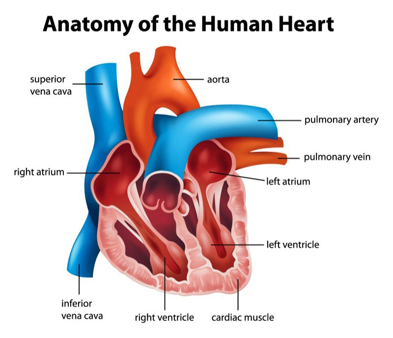
Let’s take a walk through the heart, following movement of blood to the lungs, back to the heart, and then out to the rest of the body.
It may be helpful to take detailed notes because you will be modeling this process for the next journal assignment.
Are you feeling comfortable with basic heart structures and the flow of blood through the heart?
Try labeling the following on this poster: right & left atrium, right & left ventricle; superior & inferior vena cava, ascending & descending aorta, pulmonary artery & pulmonary vein, oxygenated & deoxygenated blood, heart valves.
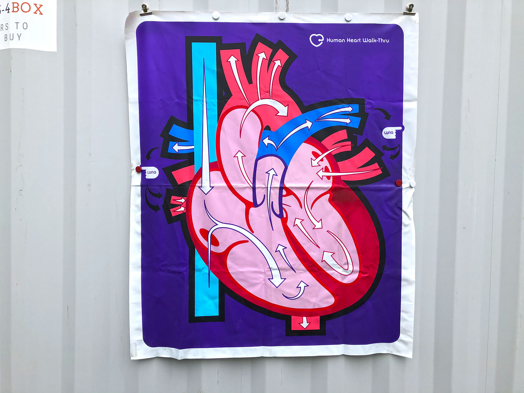
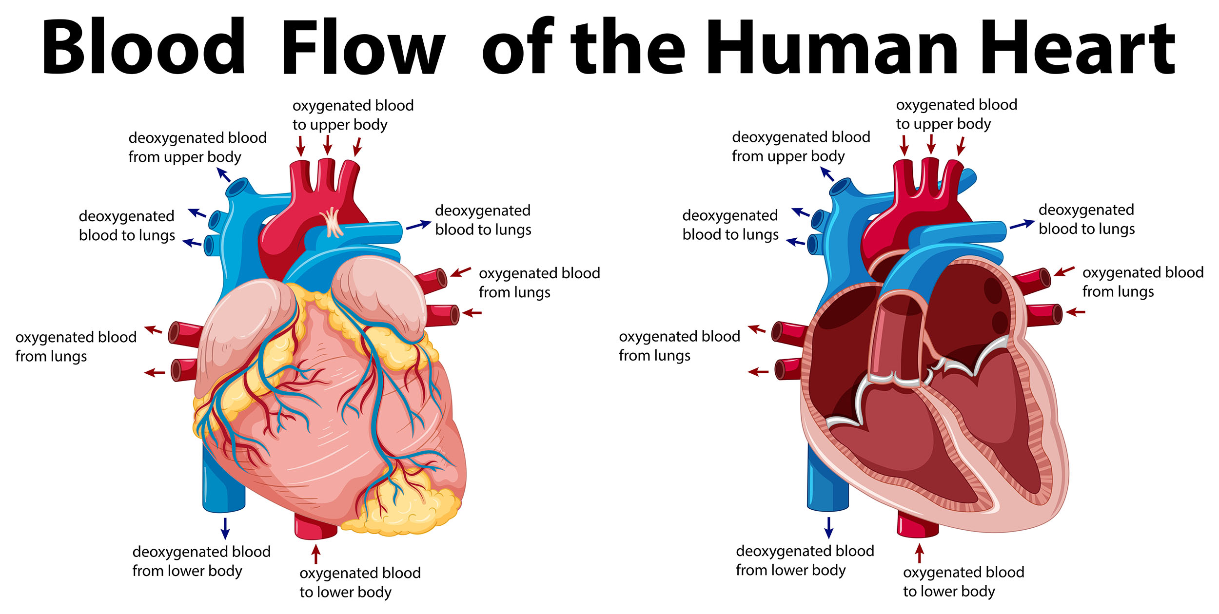
This illustration shows some of the heart structures and arrows to indicate blood flow, but a static image like this may be challenging to study.
For the next journal assignment, you will be creating your own model that teaches about heart anatomy and circulation of blood through the heart.
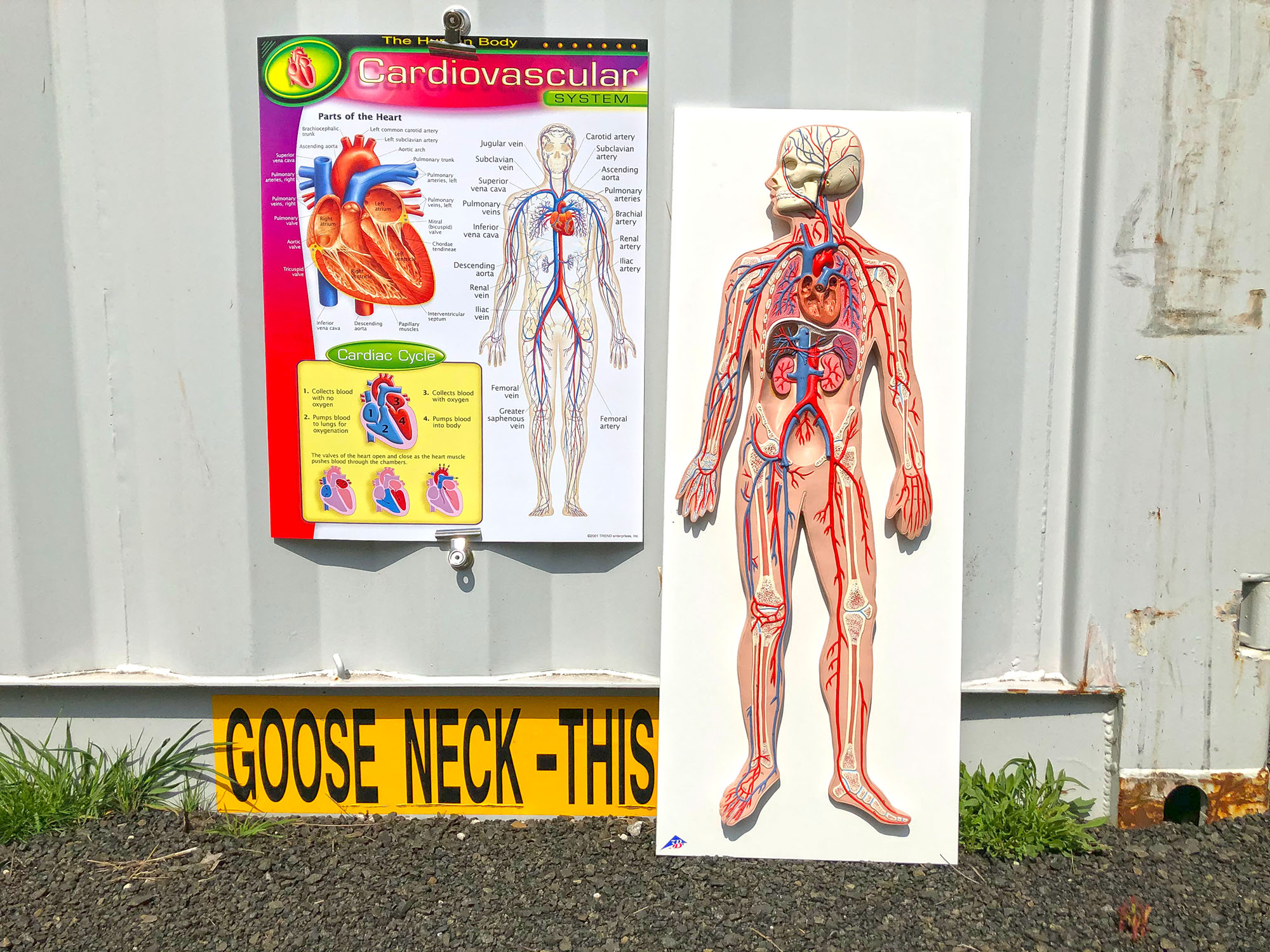
In the cardiovascular system, focus is often placed on the heart that creates pressure and pumps the blood. The vessels are also critical; these tubes carry blood throughout the body, even feeding the heart itself.
We will start with an overview of vessels, examine different types of blood vessels, and then learn about measuring pressure in arteries.
Arteries and veins are under significantly different pressures. Which one is under the highest pressure?
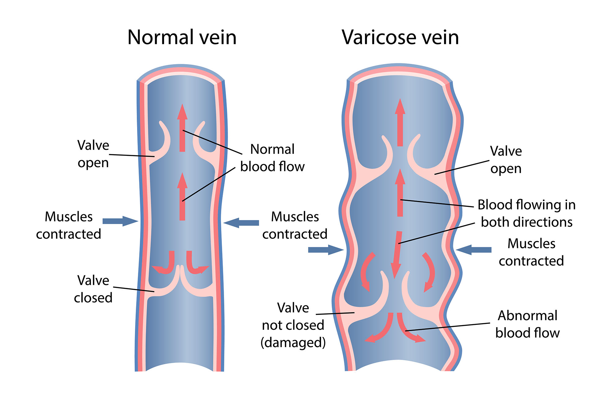
The valves in some veins can fail over time, leading to a condition called varicose veins. This can cause cosmetic changes of more visible veins near the surface of the skin, or more medically serious disruptions in blood “back-flow” in larger veins. Causes vary, and can include age, genetics, hormones, and injury.
Blood Pressure
All of us will have blood pressure measured many times over our lifetime. This is a demo on how to monitor BP at home, including what is happening to arteries during this test.
We’ll start with an overview of basic blood components.
Blood cells are tiny, so we use models, posters, and microscopic specimens to study normal blood characteristics.
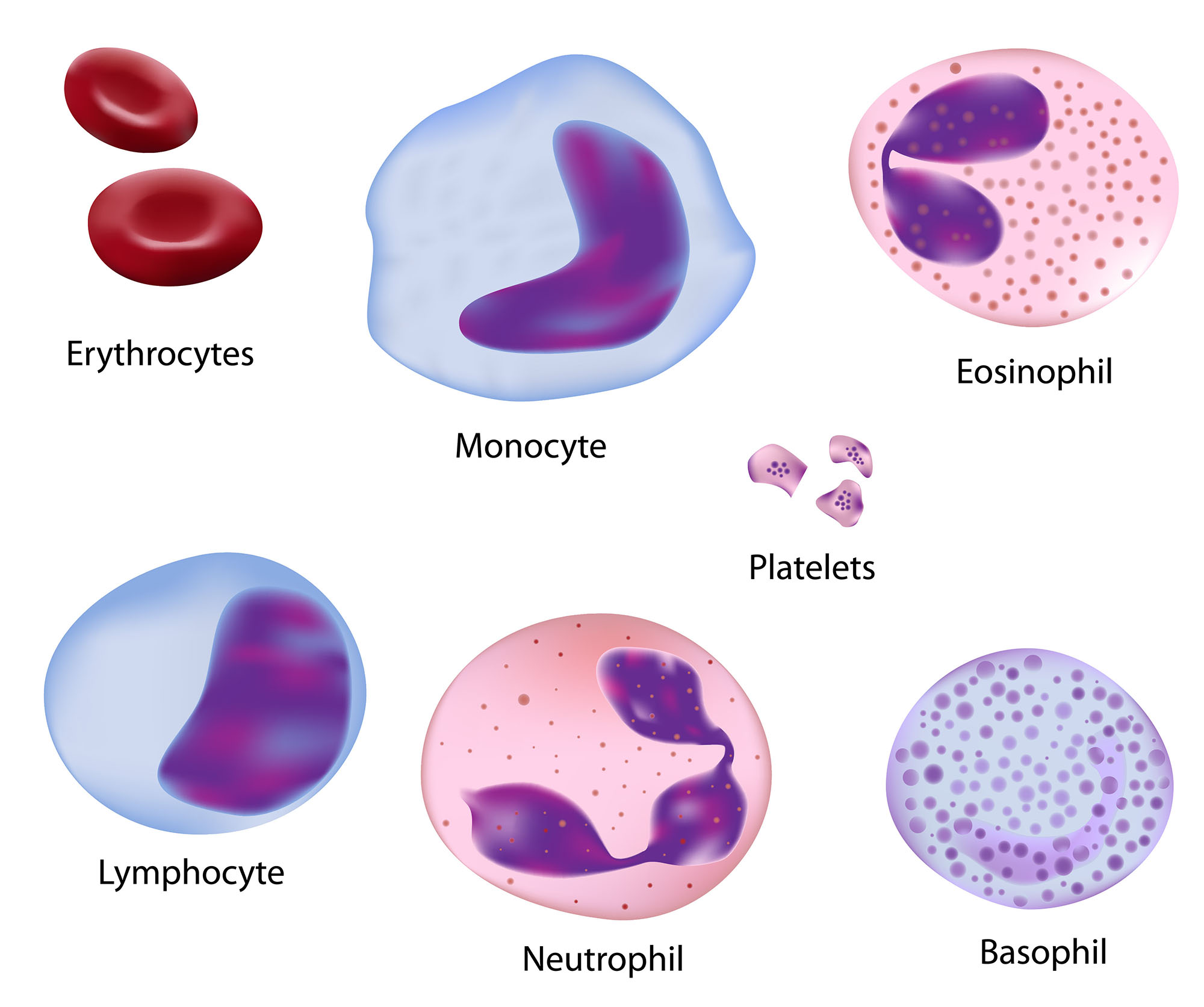
In this guide we are learning what the five WBCs (white blood cells) are; we will focus on what they specifically do in a later guide on our Defense Systems. At that point you will be using the notes you take today on WBC features.
Here is a gallery of blood components to supplement your notes. There are hints of white blood cell functions; there is more information on this in an upcoming guide.
Blood Components
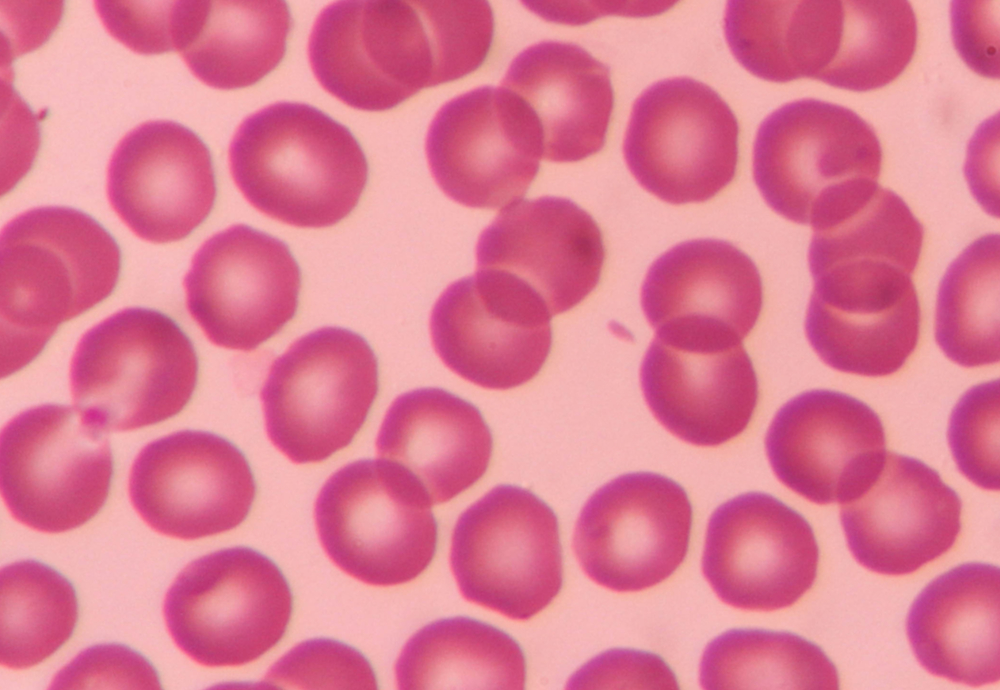
Erythrocyte
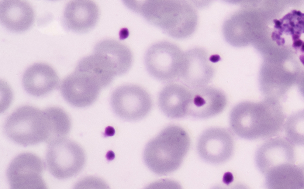
Platelet
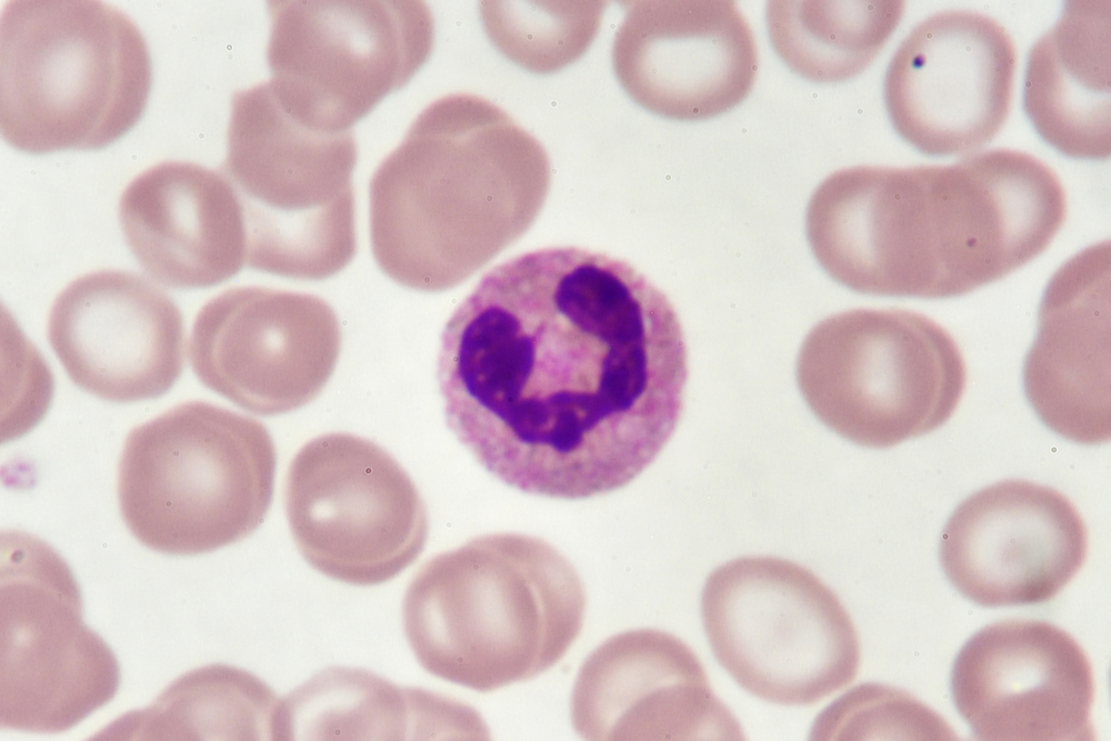
Neutrophil
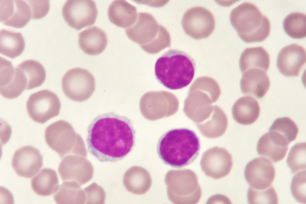
Lymphocyte
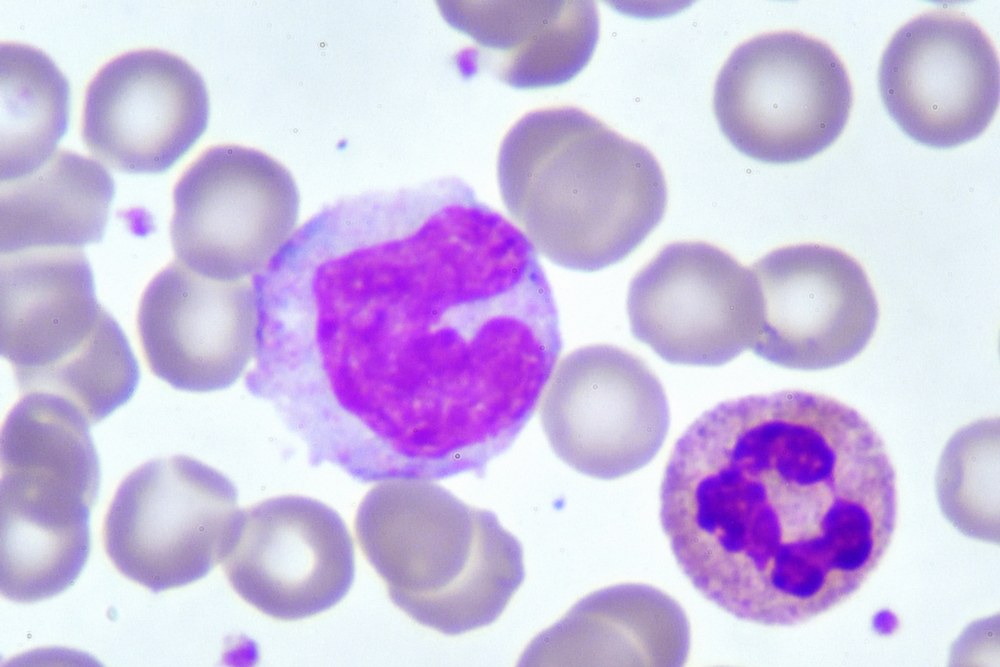
Monocyte
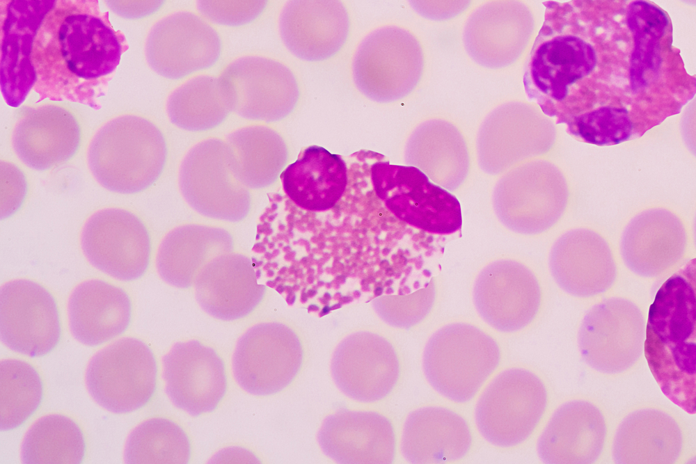
Eosinophil
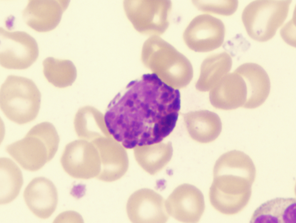
Basophil
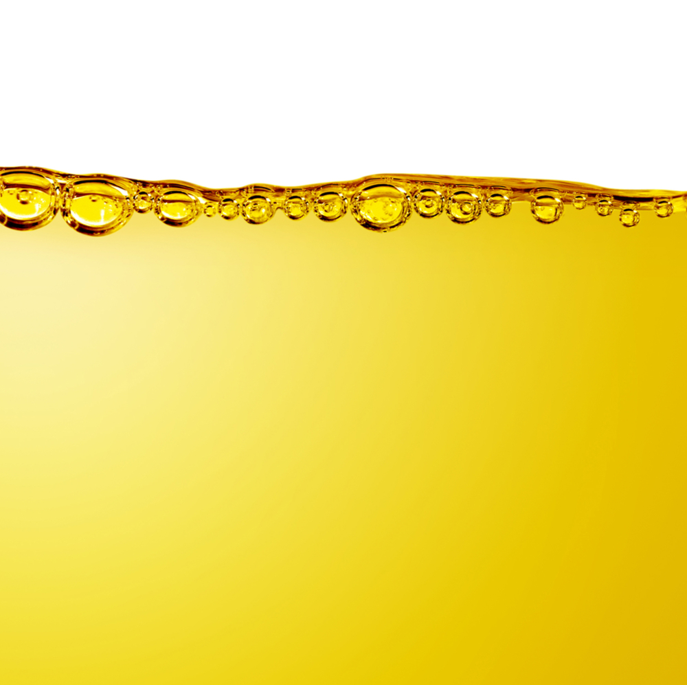
Plasma
Start Journal Assignment #4 Here
Journal Page #4: Heart Model
For this journal page you are making a model of the heart that teaches basic heart anatomy and shows how blood circulates through the heart. This is your original work, we are providing ideas, and you are making your own heart.
Your model should include: oxygenated blood, deoxygenated blood, various vessels (vena cava, pulmonary artery, pulmonary vein, aorta), lungs, and heart (all four chambers, valves).
You model could be a drawing, puzzle pieces, a digital animation, a poster, a clay model, or other creative representation.
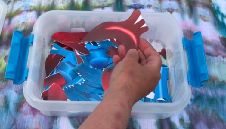
You are turning in:
A journal page with evidence of the model you have constructed, including information on how it functions. This could include a labeled sketch of the model, a series of photos, a photo and detailed description, a video showing the model, or other representation. If the journal assignment file is very large, like a long video, you may want to host it on a website to reduce upload time. Your model should demonstrate basic heart anatomy and show how blood circulates through the heart.
You can involve family members or housemates in constructing and/or demonstrating the model, if desired. Be creative and have fun creating your model. This could be an excellent piece to add to your final portfolio.
There are additional examples of possible heart models on this guide’s resource page.
The next section explores various cardiovascular disorders.
Check your knowledge. Can you:
- label the structures of the heart, including the chambers, coronary vessels, and cardiac cells and explain what occurs in a “heart-beat?”
- describe the structures and functions of arteries, veins, and capillaries and their relationship to blood pressure?
- list the various blood components, including the relative amounts and roles of red blood cells (RBCs), white blood cells (WBCs), and platelets?

