
Skeletal System Support, Protection, & more

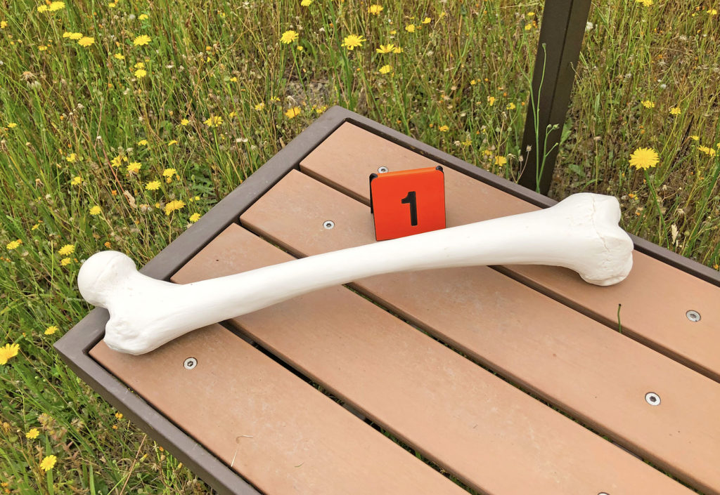
Skeletal System Objectives
- Provide examples of different organ systems, including the hierarchy of organ system, organ(s), tissues, and cells.
- Describe skeletal system structures and functions, including the system’s organs, tissues, and cells.
- Identify various bone disorders, including the organ that is impacted.
Start Journal Assignment #3 Here
Journal Page #3: Quiz Analysis
Now that you have taken the 1A quiz and are beginning material for the 1B quiz, it is a good time to reflect on studying and quiz preparation.
Answer the following questions on a journal page. You do not need to include the questions, only your responses to each question. You can include additional relevant information if you like. Simply writing the responses may inspire new study strategies.

Provide thorough and complete answers to each of these questions on your quiz analysis journal page:
- Did I recognize all/most of the material on the quiz? ___________ (can indicate whether studying covered the full range of material)
- Did I think I would perform better on the exam than I actually did? ___________ (indicates learning to recognize material rather than retrieving answers)
- Were there multiple answers I changed, or wasn’t sure about? ____________ (if correct answers don’t pop into the mind, studying may address this)
- Did any particular source of material seem to cause more difficulty than other sources? (text or videos?)
- What was my study strategy for this quiz?
(time spent, study locations, study materials made, etc.) - What are two possible ways to improve studying and learning material for the next quiz?
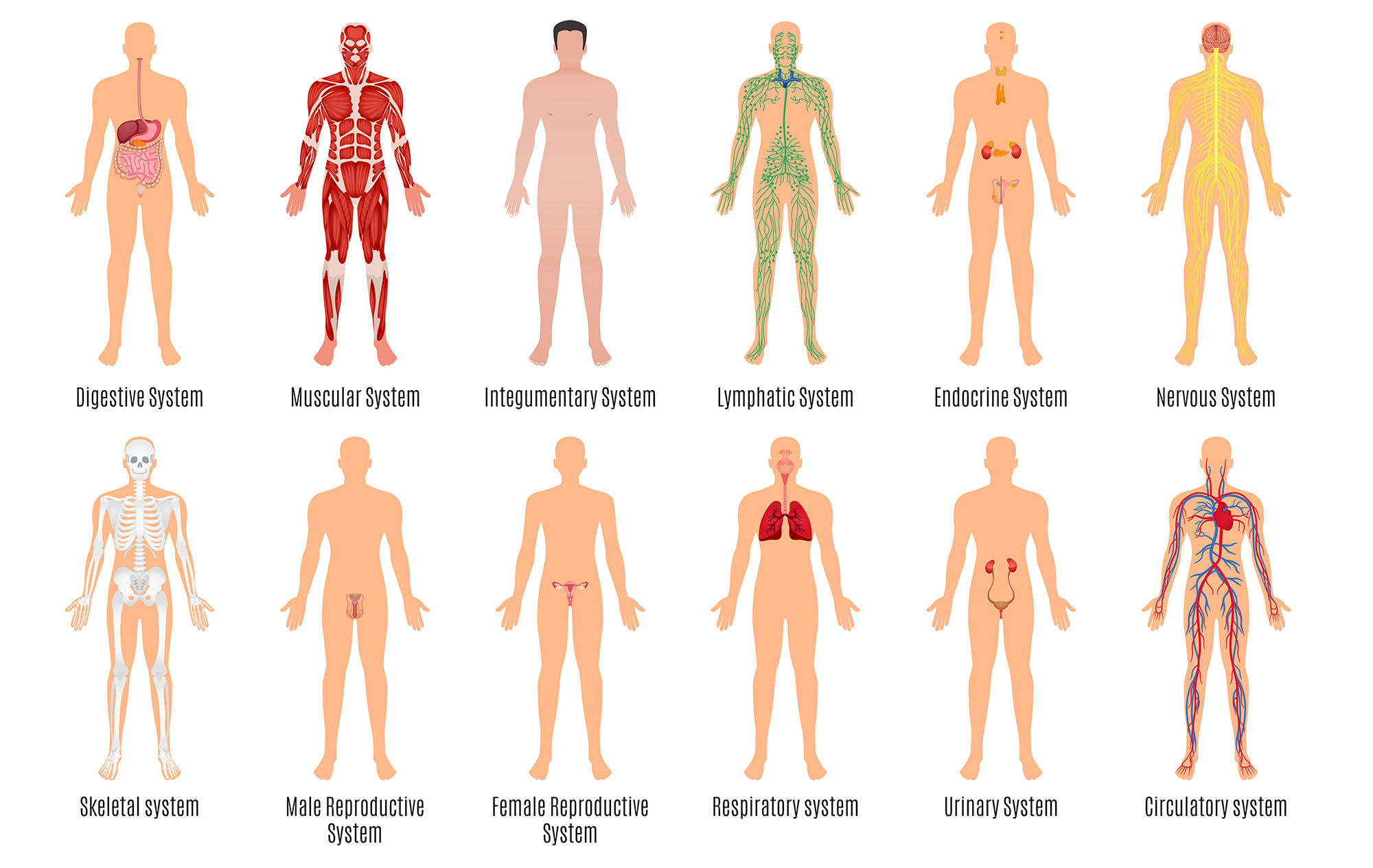
One of the earliest things we learn about the human body is that we have different organ systems made up of organs. For example, the urinary system has kidneys and a bladder; the cardiovascular system includes the heart and blood vessels, and the skeletal system is made up of bones.
Organ systems are made up of organs, and many of those organs are generally familiar. What is less familiar is that organs are made up of tissues, and tissues are groups of cells.
The integumentary system we covered in the last Guide was pretty straight forward, an organ system with one basic organs (skin) and it’s appendages (hair and nails). The skin organ is made of tissues: epithelial in the epidermis, loose connective in the dermis, and adipose connective in the hypodermis. We also met some of the cells in the tissues: keratinocytes in the epithelial tissue of the epidermis, fibroblasts in the connective tissue of the dermis, and adipocytes in the connective tissue of the hypodermis.

Organ System
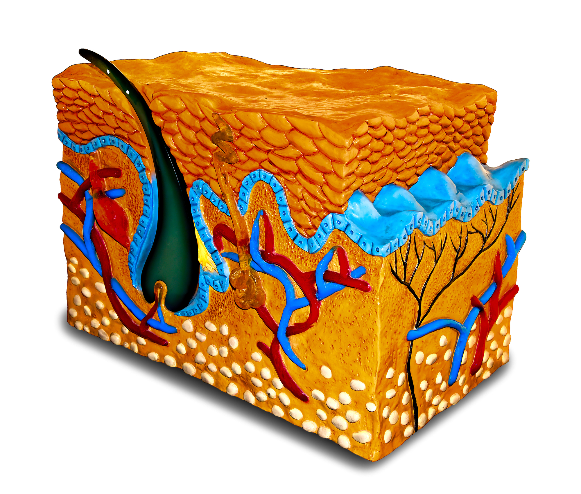
Organ
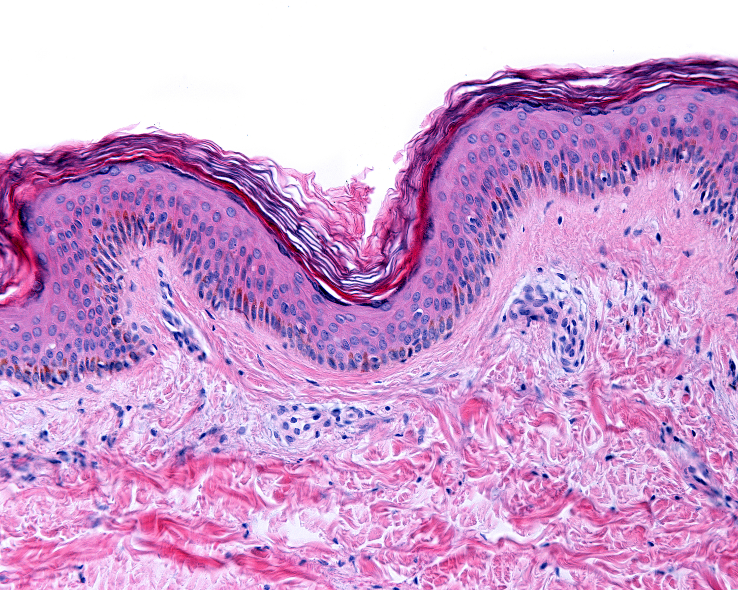
Tissues
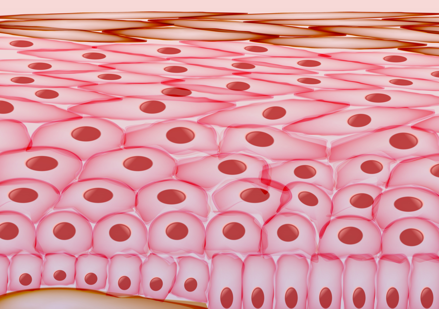
Cells
We will be building on the basics you learned in the previous Guide. For example, we will be back to skin when we discuss the impacts of aging on organs.
Check your memory of skin basics with this video.
The skeletal and muscular systems are also straight forward. This video explains why.
This video uses an inexpensive flip chart to provide an overview of the organ systems we are covering in the course.
Skeletal System
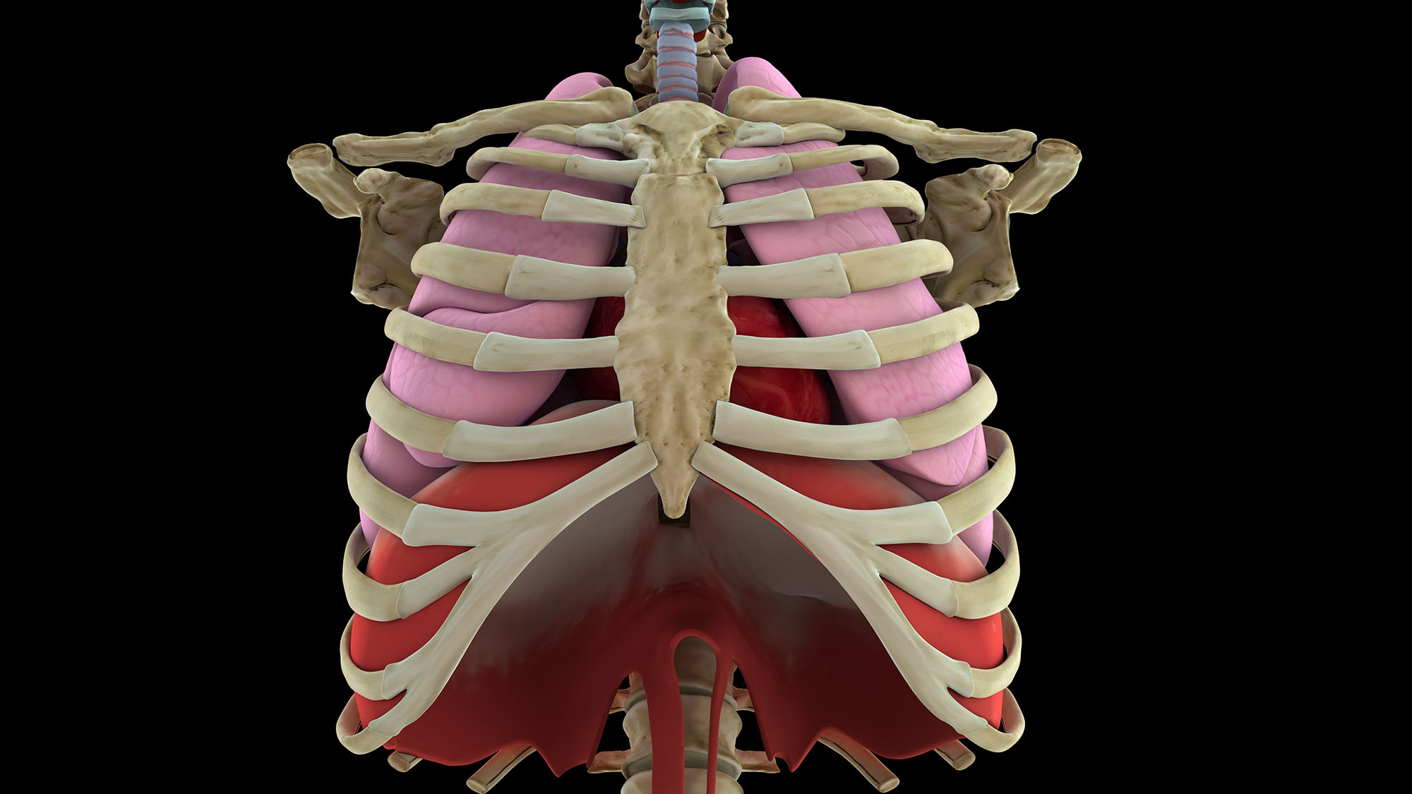
The skeletal system and its bone organs has several basic functions. How many can you list?
The video below introduces the skeleton’s basic functions and supporting structures.
This video introduces the specific bones you need to know for this course.
Need to see this poster further? There is an additional video (in a hailstorm) on this guide’s resource page.
The human skull may be the most recognizable part of the human skeleton. Our skull includes a cranial vault protecting the brain and elaborate facial bones. The hinged jaw is split into two parts: the upper maxilla, and the lower mandible.
The human backbone needs to be strong enough to support our large head and upright posture, as well as flexible enough to allow a wide range of movements.
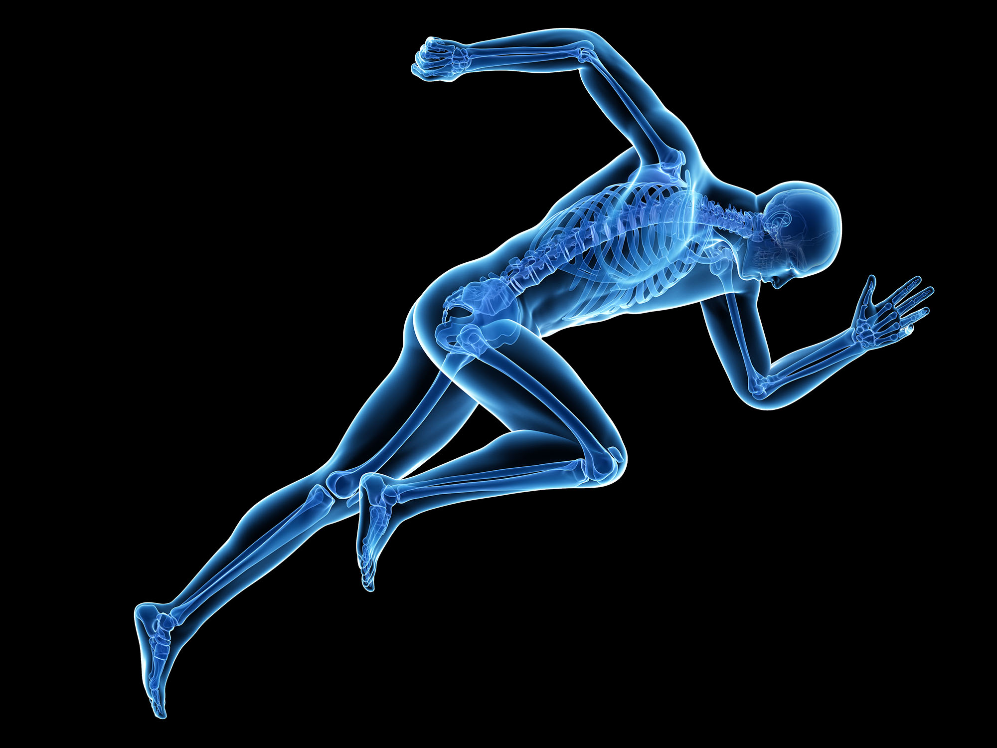
Unfortunately, many people develop back injuries over time. Besides chronic (recurring, long-term) headaches/migraines, chronic back pain is the leading cause of pain worldwide. People often mention a “cervical injury” or “lumbar pain,” this video shows where these occur on our spine.
Back pain can be due to small fractures in the vertebral bones, but it can also be caused by damage to the disc padding between the vertebrae. This has a variety of names, including a herniated disc, a slipped disc, or a disc prolapse.
If you have to pick up something heavy, and it’s really best not to, at least you can try to minimize impact on the spine. Mark demonstrates how.
Bone Structure
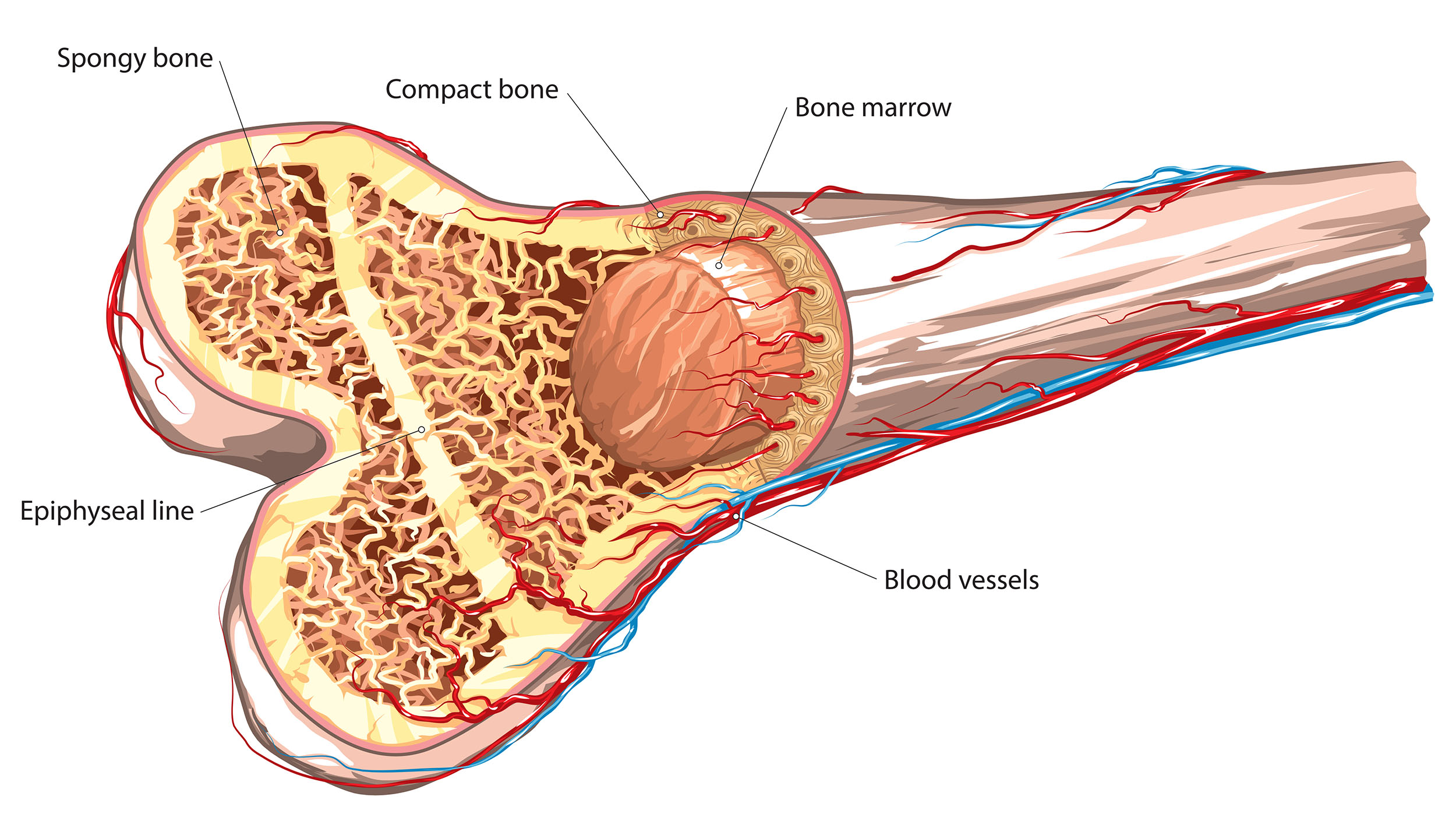
Bone architecture often has spongy bone on the ends and compact bone in the middle. Spongy bone is more flexible and less likely to break than compact bone. It has other important attributes discussed in the next video.
Now we are taking a closer look at bone tissues and cells.
Bone is continually remodeling based on changes in our activities. This image is of osteoblasts (purple) on the outer layer of bone (pink). These cells can extract calcium and other minerals from the blood and deposit it in a bony matrix.
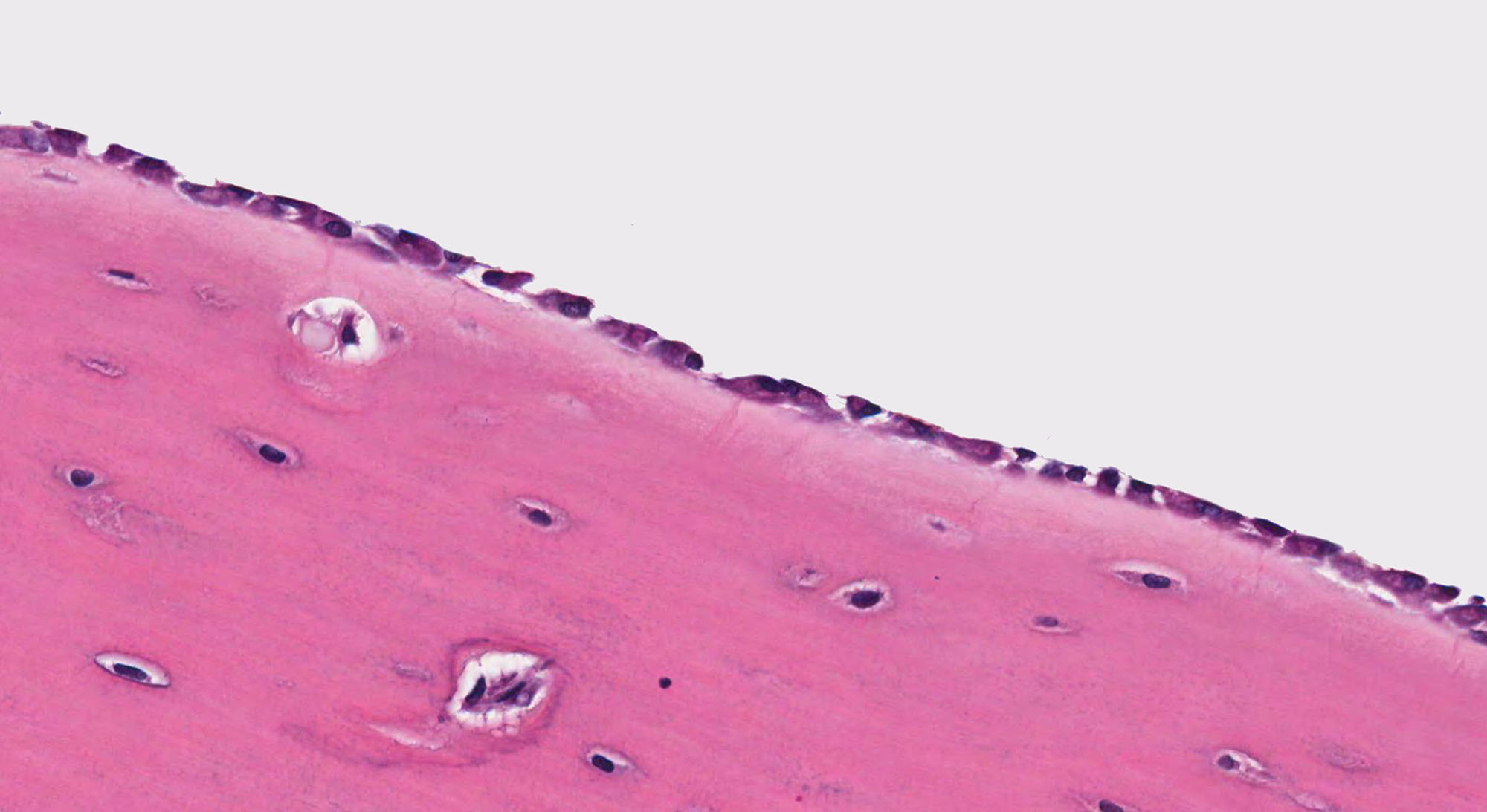
Osteoblasts build bone, and osteoclasts do the opposite. Why is this balance important?
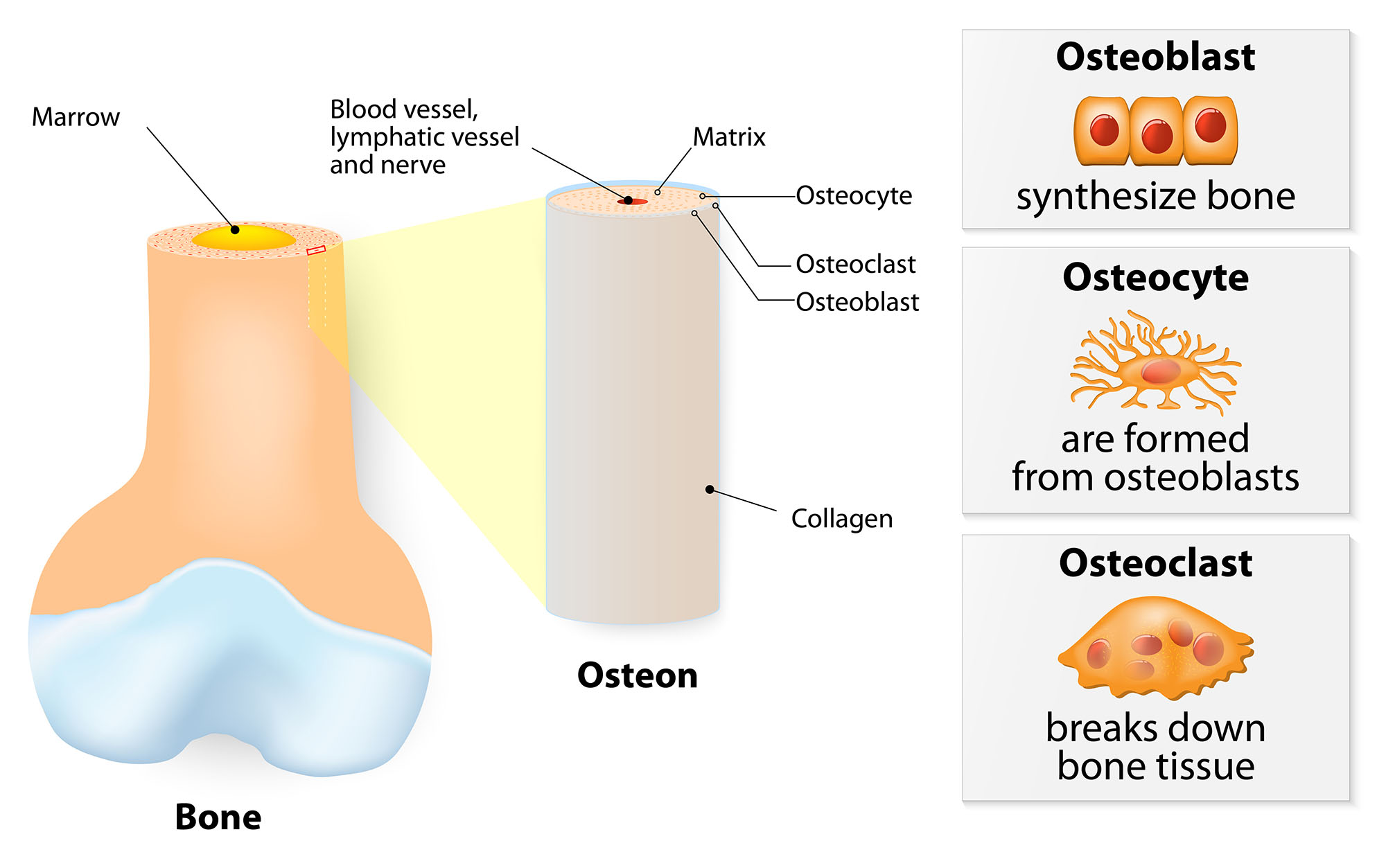
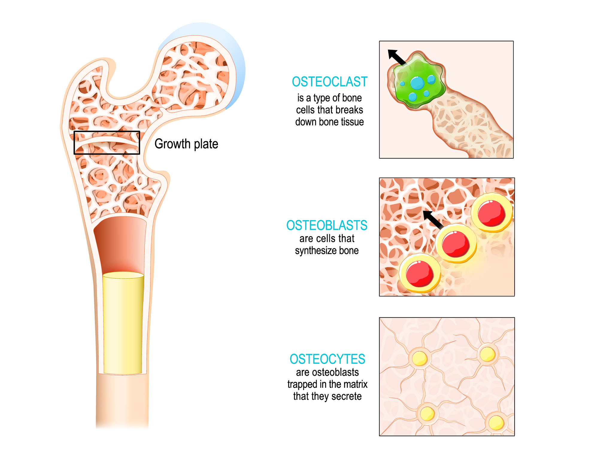
Let’s examine bone under the microscope.
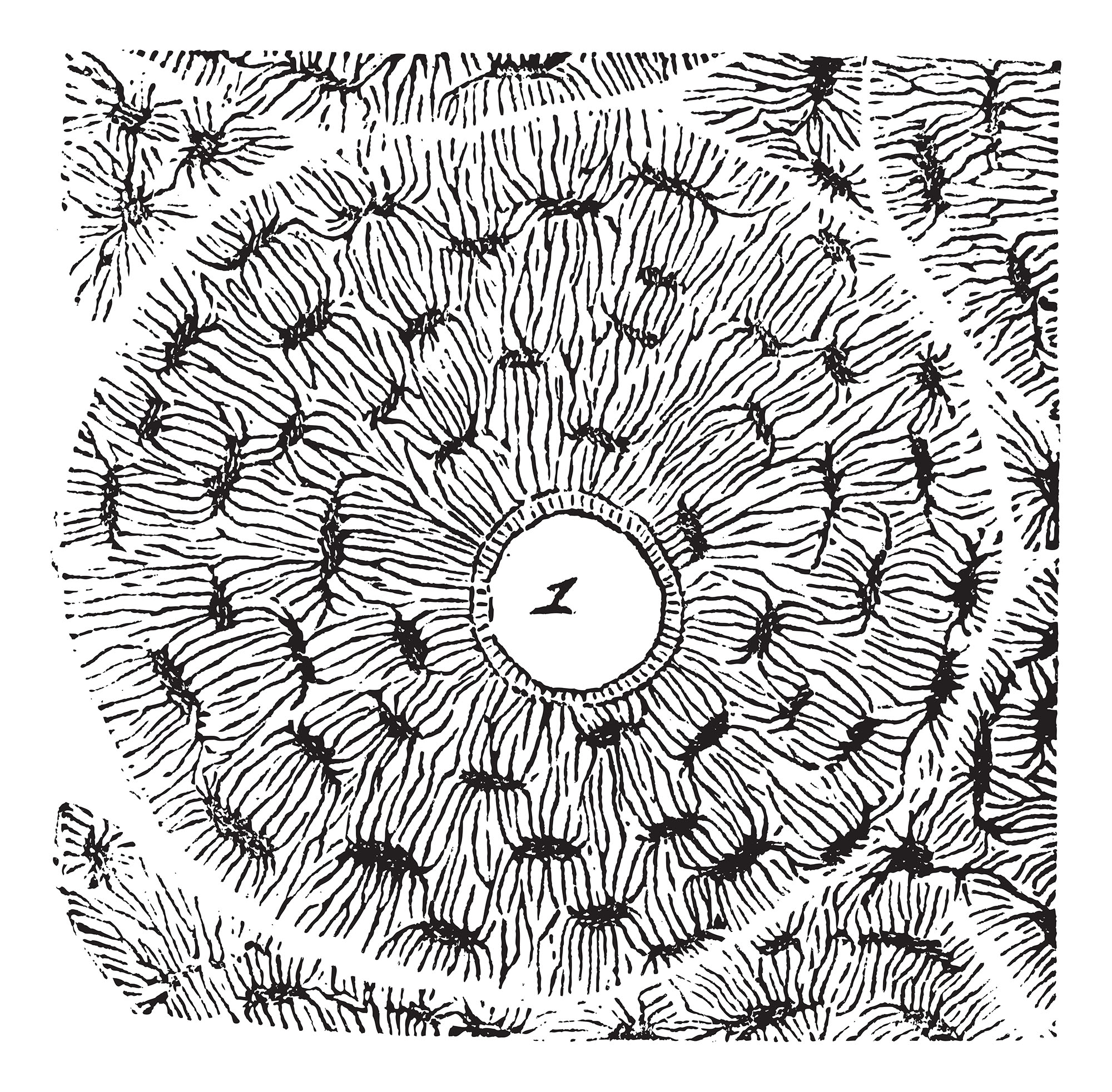
In this figure,
What runs through the center of the osteon (labeled #1)?
What are the living cells cemented within the bone?
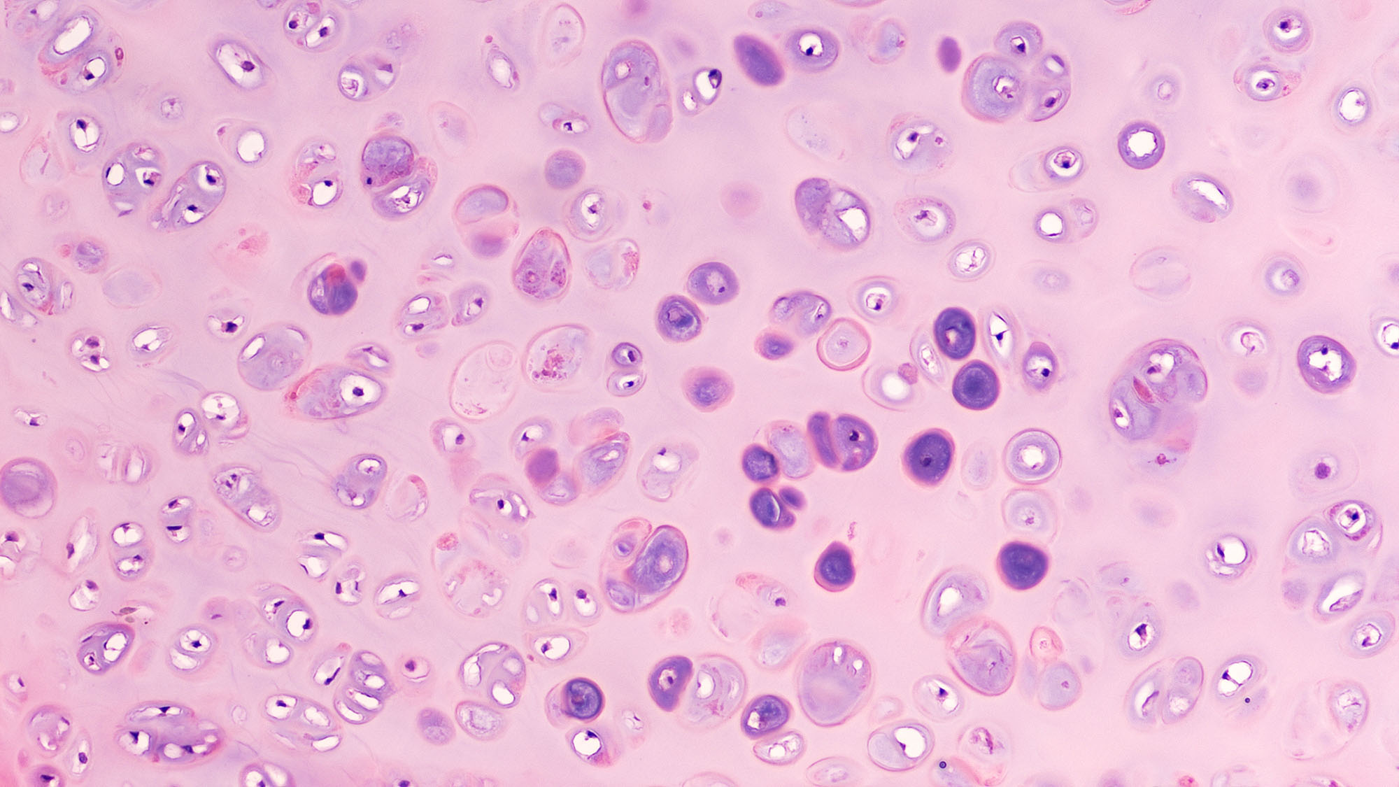
In the microscopy video above, the first image is of a disc between vertebrae. Discs have a different type of connective tissue than bone; cartilage. Cartilage is found in many firm but flexible structures like your ears or padding between bones. The cells (chondrocytes) have a unique appearance, like there is empty space around them. We’ll look at them in closer detail when we cover the respiratory system because cartilage also lines the outside of the trachea (windpipe).
Joints
Bones come together in joints. This video introduces the various joints you need to know for this course and may injure in your lifetime.
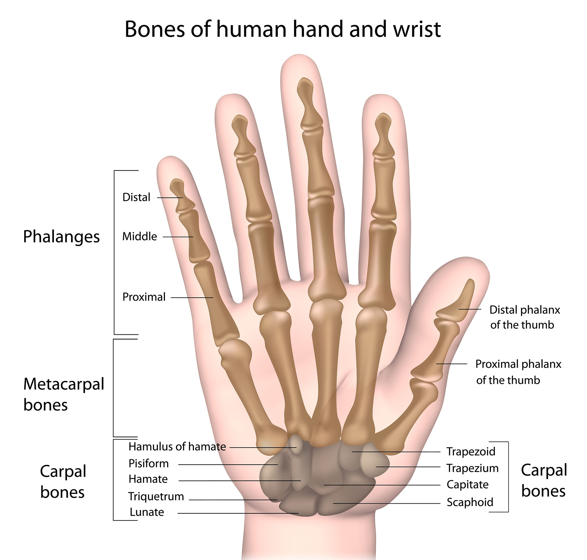
The most elaborate joints are in our hands and feet. This video introduces the bones; keep in mid all of the possible sites of injury.
This image shows bones of the fingers, palm, and wrist. Which sole bones in the foot are equivalent to the carpal bones in the hand? Which ankle bones in the foot are equivalent to the metacarpals in the hand?
Bone Disorders
Most of us will have multiple skeletal fractures in our lifetimes and approximately half of us will develop inflammation disorders, including arthritis. This video introduces these disorders.
Fractures are typically diagnosed with X-rays. You have likely experienced a medical professional showing you an X-ray to explain a diagnosis. This video introduces how to read an X-ray.
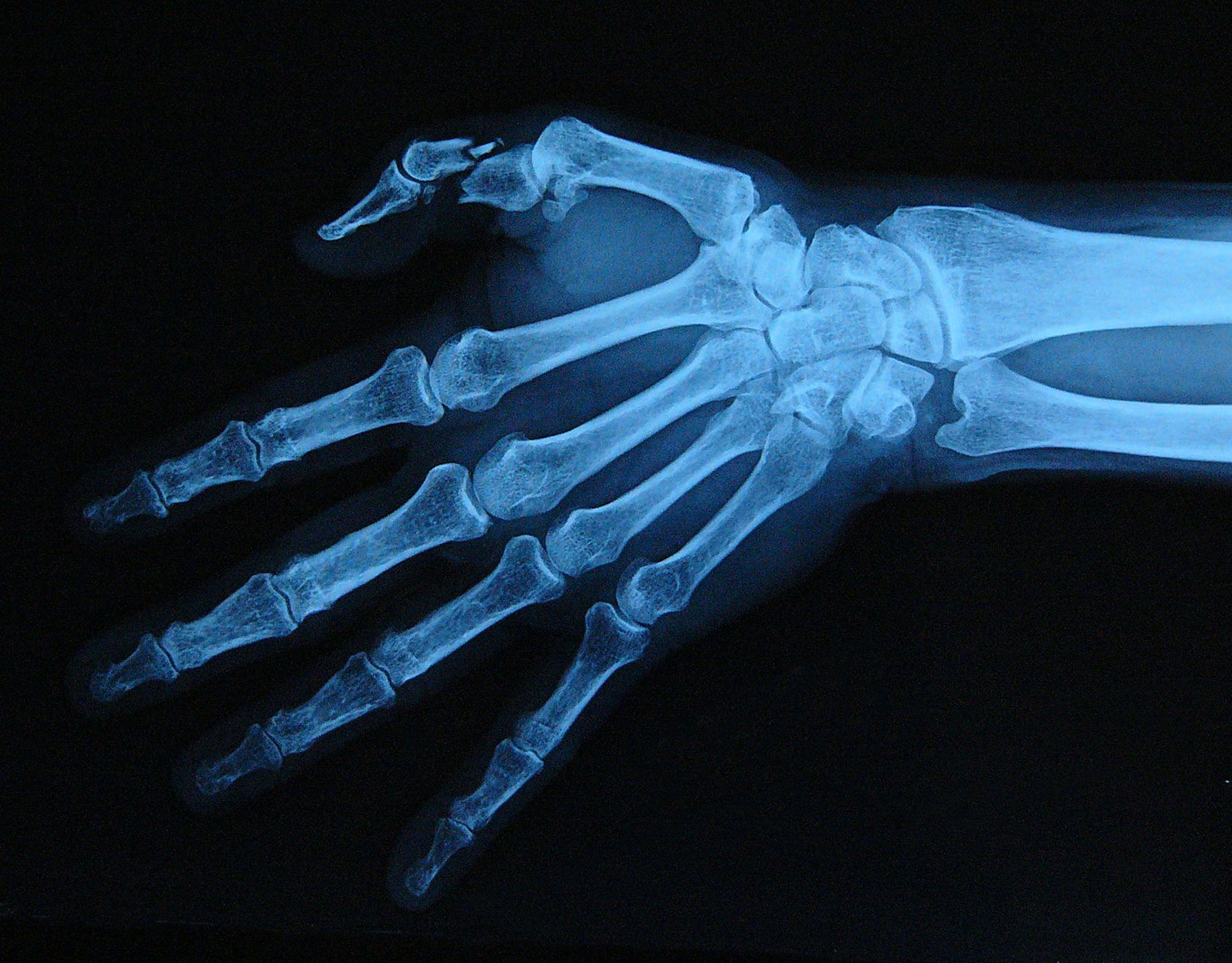
1. Broken thumb
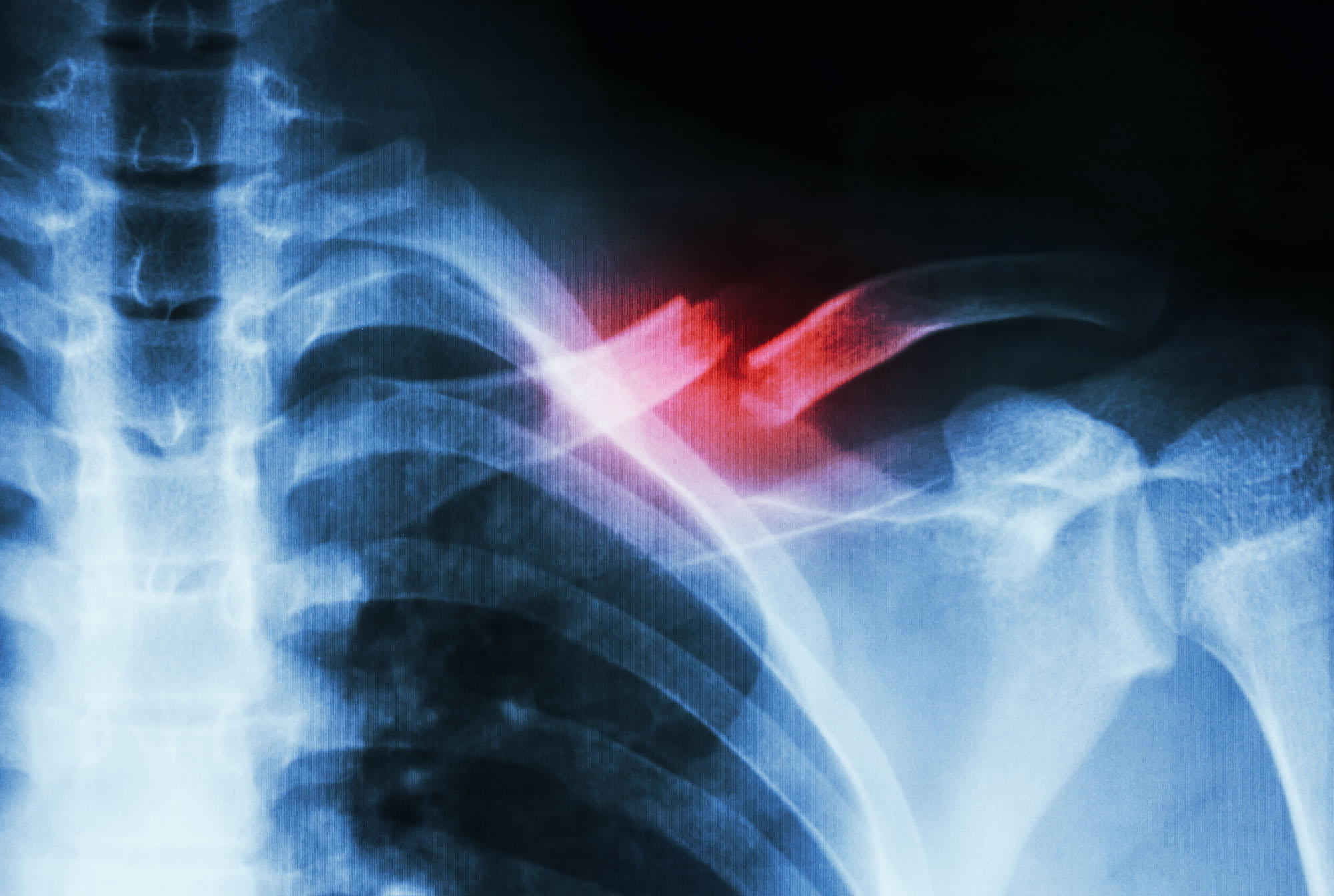
2. Broken clavicle
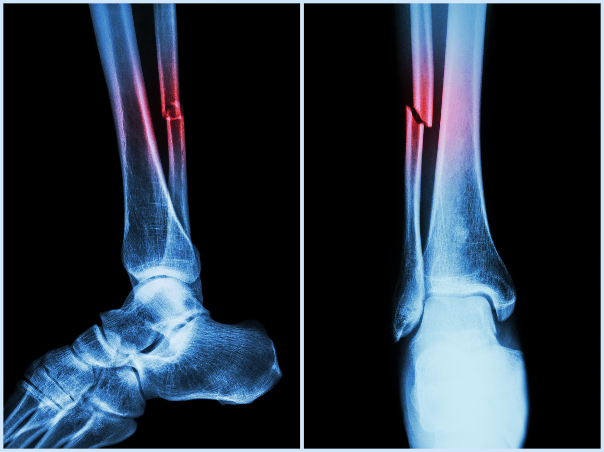
3. Broken fibula
Often a medical professional will identify the type of fracture. Here are the most common forms you may encounter.

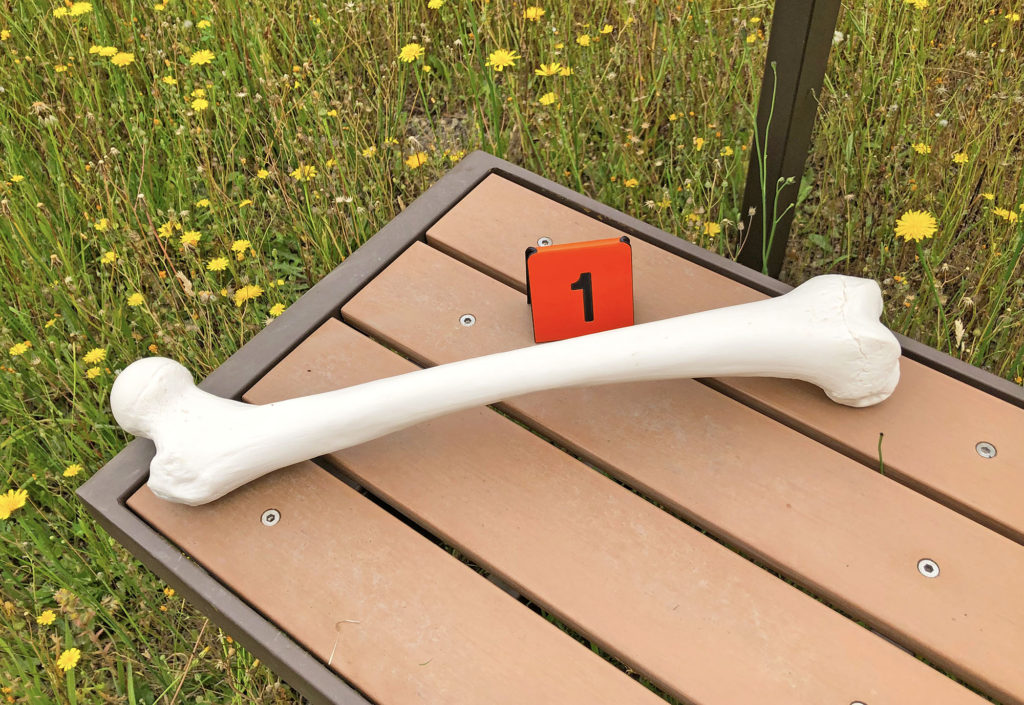
If a bone fractures it can heal within 1-2 months if it is a small break, or many months if it is a large break, like the femur of the upper leg or the tibia and fibula of the lower leg.

Arthritis
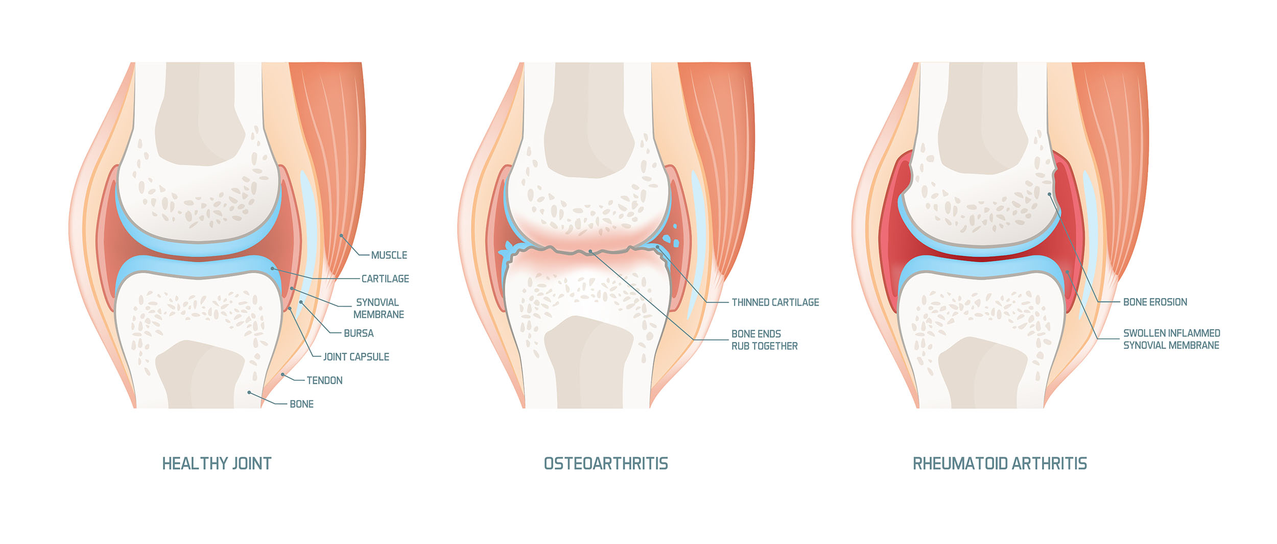
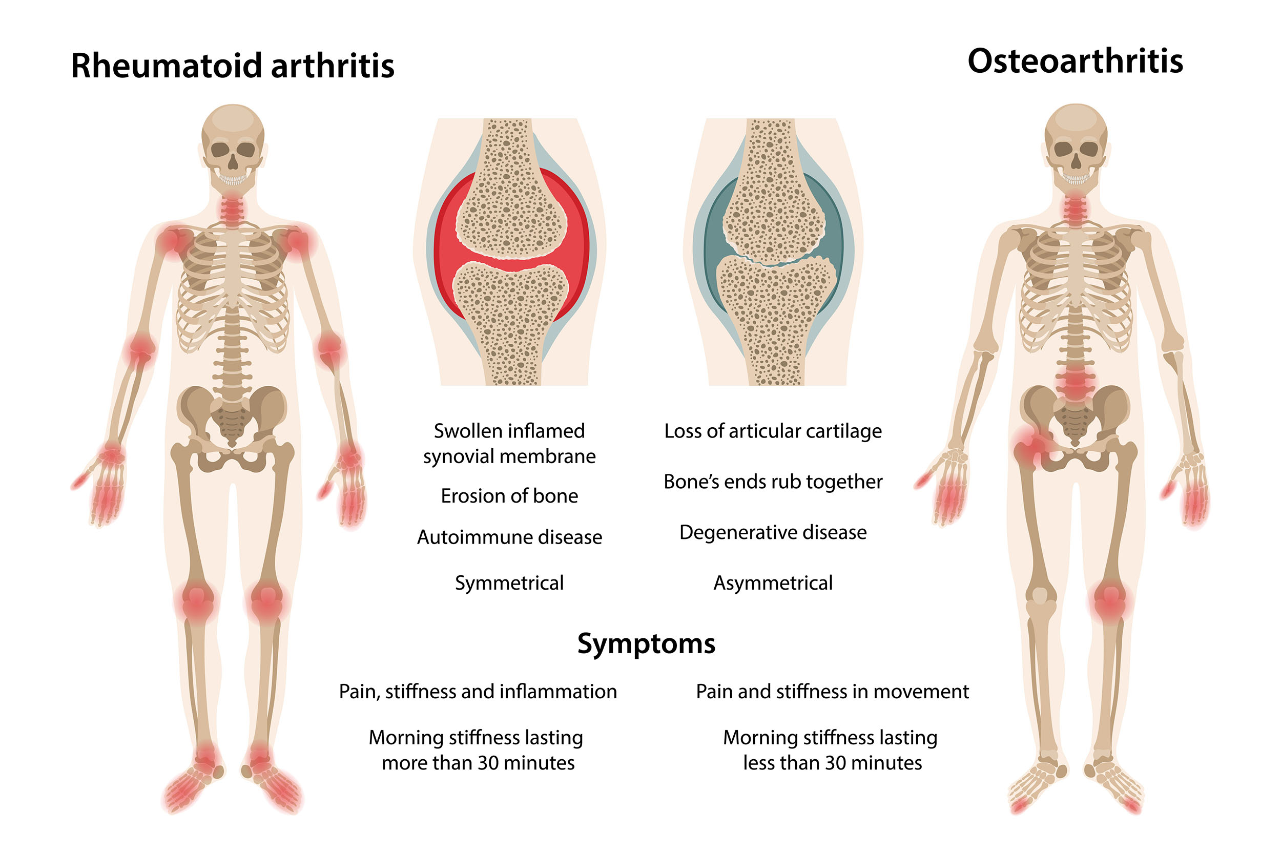
Arthritis is inflammation at a ___, where two bones come together (see above). Rheumatoid arthritis is an autoimmune disease, the body attacks its own joints instead of pathogens like viruses or bacteria. Osteoarthritis results when padding at joints deteriorates and bones are displaces and/or grind together.
Bone density can decrease with age; osteoporosis is a disease that speeds up the rate of bone loss.
Osteoporosis can go undetected until a fracture occurs. The most dangerous can be a “hip” fracture because it immobilizes an individual and takes a long time to heal. Healing is further delayed if a person is older and has limited calcium absorption.
The next section introduces the muscular system and how it interacts with the skeletal system.
Check your knowledge. Can you:
- provide examples of different organ systems, including the hierarchy of organ system, organ(s), tissues, and cells?
- describe skeletal system structures and functions, including the system’s organs, tissues, and cells?
- identify various bone disorders, including the organ that is impacted?
